Mast Cell Tumor Cat Cytology
Mast Cell Tumor Cat Cytology - Cat Meme Stock Pictures and Photos
aniDAP Cytology>Mast cell tumor

Mast cell courtesy Dr. Undhad Vishal Brookfield Animal Hospital

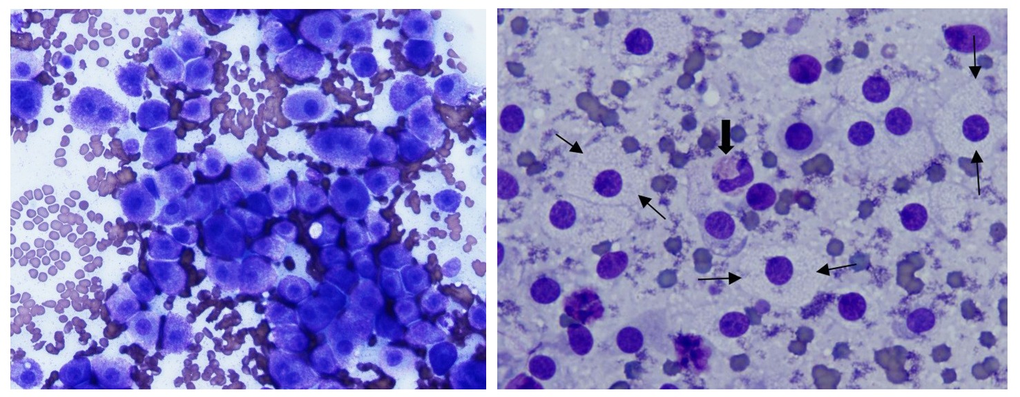
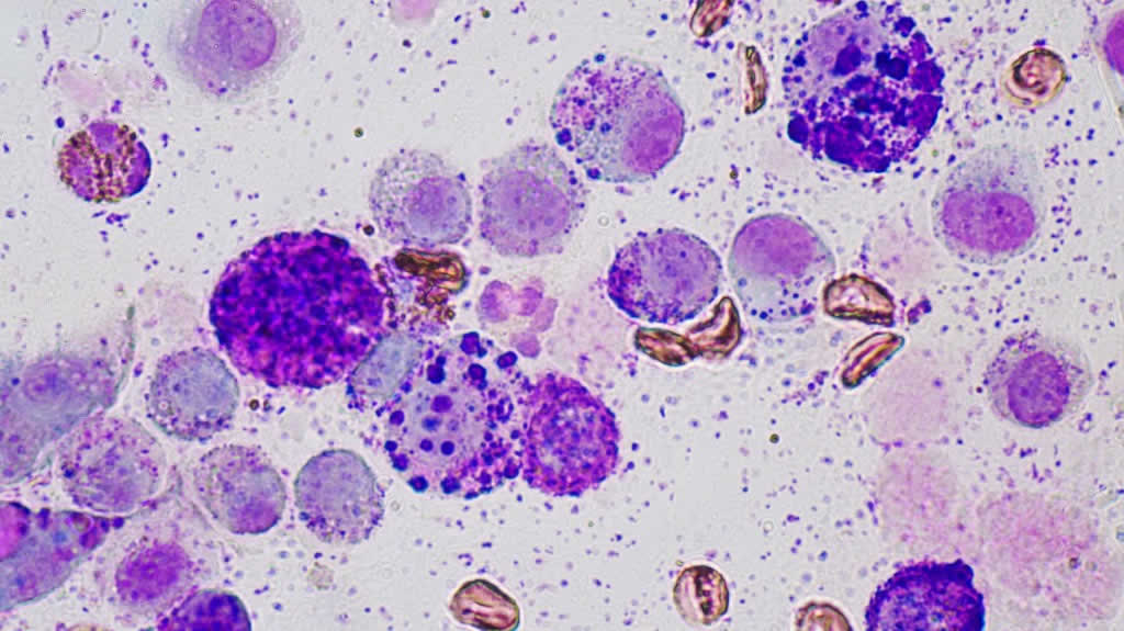
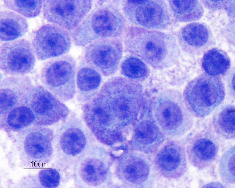
Poorly differentiated mast cell tumor FNA cytology from a dog. Eğitim
Color Cytology Atlas of Common Feline Skin Tumors • MSPCAAngell

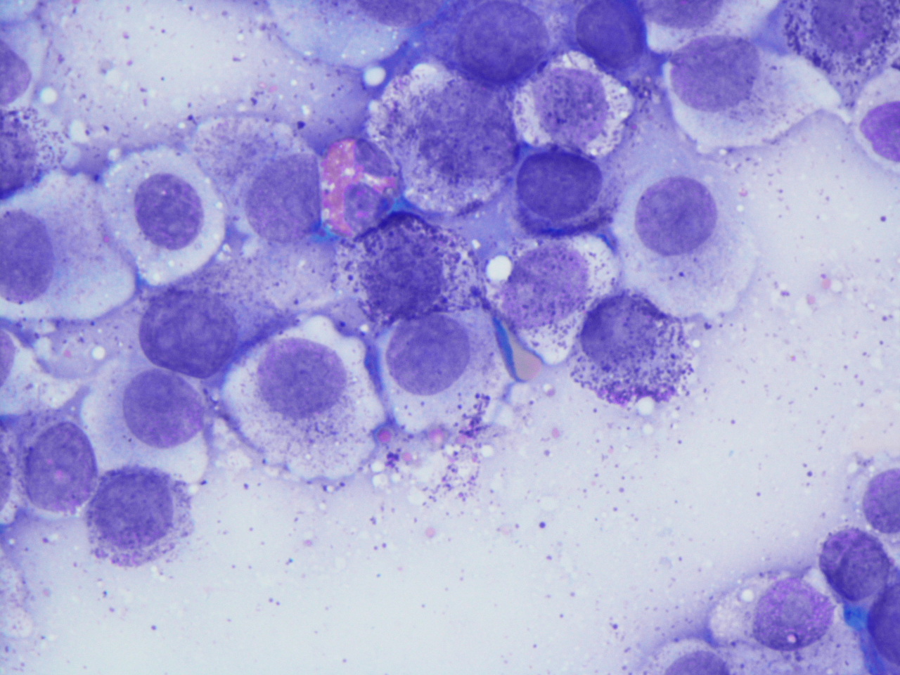
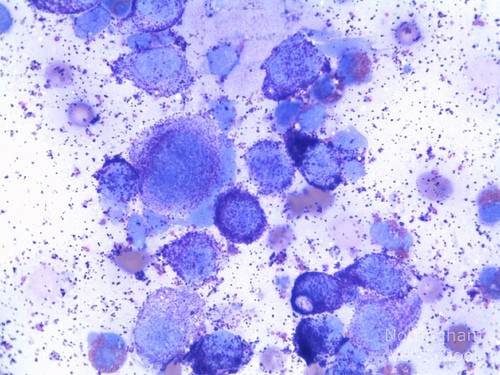
"Discrete" cell tumors eClinpath
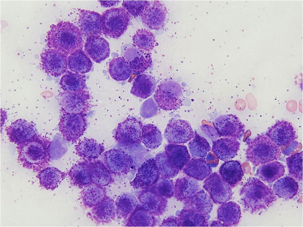
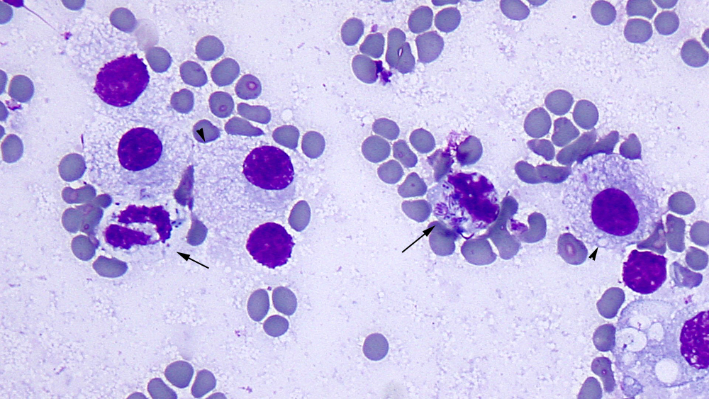
Canine Mast Cell Tumours

They are usually noticed in middle aged patients, but can occur in patients of any age.
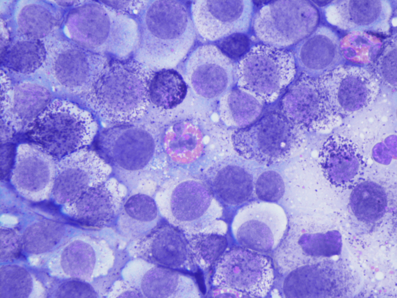
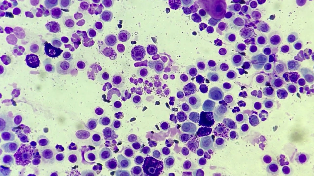
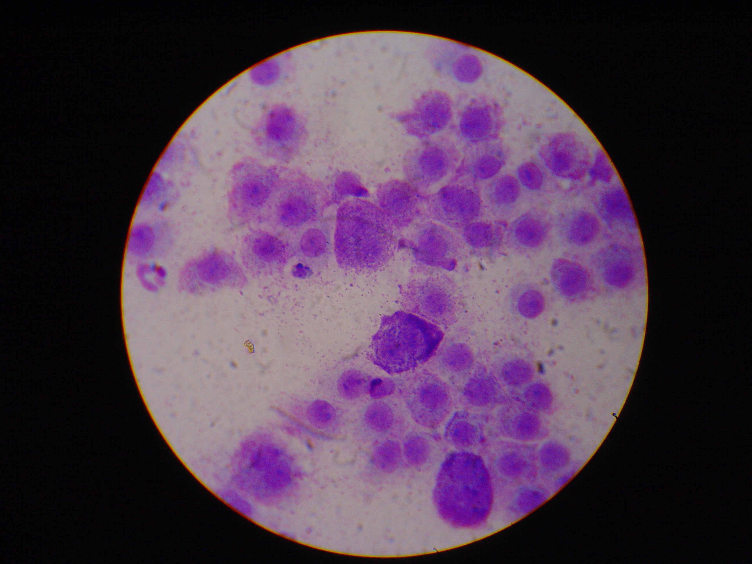
Mast cell tumor cat cytology. Mast cell tumors (mct) represent a cancer of mast cells, which are a type of white blood cell involved in allergy and inflammation. Veterinarians classify mast cell tumors (mct) anatomically as being either cutaneous (involving the skin), or visceral (involving the internal organs). Approximately 20% of skin masses in cats are cutaneous mast cell tumors, and about 90% of those are benign.
Intestinal mast cell tumor in the cat is distinct from systemic mastocytosis (which does not involve the bowel). The masses are usually pink and alopecic, and they may be smooth or ulcerated. Pathologists divide mast cell tumors into two forms:
Cytology will be carried out these fine needle aspiration samples, with a pathologist examining the samples, checking for high levels of mast cells. Mast cell tumors typically affect older cats; They are small, firm, raised, hairless and can become itchy.
Mast cell tumors (mcts) arise from malignantly transformed mast cells. Mast cell tumours commonly affect the skin, the spleen (in the abdomen) and/or the intestines. The algorithm below taken from the original publication shows the criteria used for grading.
If your cat has the splenic form of the disease, the most commonly observed signs are weight loss, vomiting,. Neoplasia of mast cells not only occurs most commonly in dogs and cats but also has been described in horses. Mcts are the most common splenic tumor, second most common skin tumor and third most common intestinal tumor in cats.
Mast cell tumors (mct) can occur in the skin, spleen, or gastrointestinal tract of cats. Cutaneous (skin) mast cell tumours most commonly affect the head and. Several studies suggest that siamese cats.
neoplasia Archives eClinpath
Atlas Archives Page 2 of 8 eClinpath
Poorly Differentiated mast cell tumour cytology YouTube
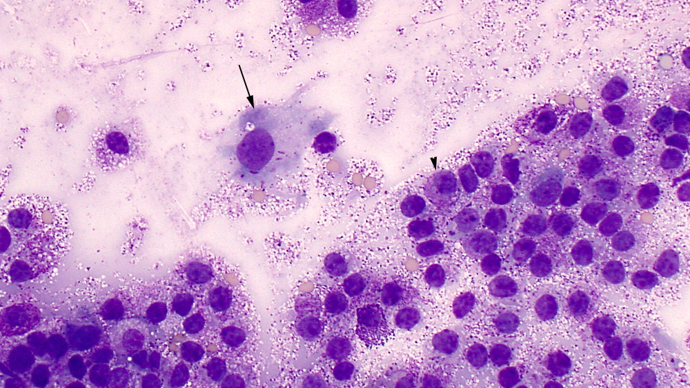
2 Cases of mast cell tumours Dermvet
aniDAP Cytology>Mast cell tumor

Cytology Common Neoplastic Skin Lesions in Dogs & Cats

Liver eClinpath

Cytology Common Neoplastic Skin Lesions in Dogs & Cats
Gastrointestinal tract eClinpath

Intestinal Mast Cell Tumor In Cats
Mast Cell Tumors in Dogs A Common Canine Skin Cancer
Mast Cell Tumours in Dogs Vet Education
"Discrete" cell tumors eClinpath
