Cat Cerebellar Hypoplasia Mri
Cat Cerebellar Hypoplasia Mri - Cat Meme Stock Pictures and Photos

Fetal MRI Cerebellar Hypoplasia

Cerebellar hypoplasia in a 16yearold patient with AT on the brain

Fetal MRI Cerebellar Hypoplasia
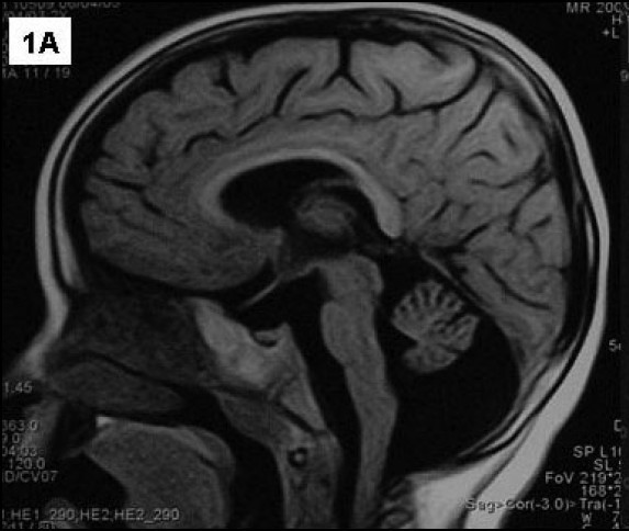
MRI Image Saggital T1 image showing hypoplasia of the inferior

Fetal MRI Cerebellar Hypoplasia

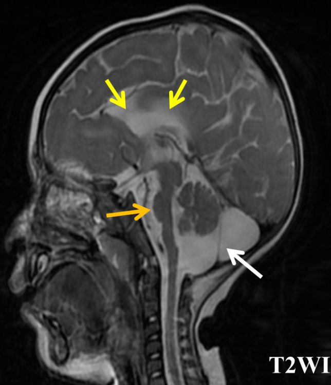
MRI of the brain indicating cerebellar atrophy. a A sagital T1 image
Unless a ch cat has other health issues, her life expectancy is the same as a cat’s without ch.
Cat cerebellar hypoplasia mri. Cerebellar hypoplasia is the incomplete development of the cerebellum. Pontocerebellar hypoplasia was first described as a specific entity by a dutch. Christopher mancuso is an active volunteer with staten island hope animal rescue and along with his wife, catherine, proud guardian of several special needs cats.
A vet or veterinary neurologist can always order an mri scan to detect an underdeveloped. Global cerebellar hypoplasia can appear indistinguishable from diffuse cerebellar atrophy on a single study and can only be distinguished from the latter by demonstrating or implying (clinically) that there has been no change over time 4. The clinical presentation is different varying from normal to severe bilateral spastic cerebral palsy, intellectual.
Cerebellar hypoplasia (cer·e·bel·lar hy·po·pla·sia) is a disorder found in cats and dogs which causes jerky movements, tremors, and generally. This disease is most common in. Your vet may also rule out other diseases by running certain tests, such as blood work.
Apprentice about the symptoms, diagnosis, and analysis if cerebellar hypoplasia in cats. Treatment and home care for cats with cerebellar hypoplasia. Since cerebellar hypoplasia is a neurological condition, there is no way to be 100% sure that your cat has ch (as opposed to another condition) unless your vet verifies it with an mri or other tests.
An mri of the brain will show a cerebellum that is much smaller than normal, and there will be more cerebrospinal fluid (csf) around it. This condition occurs in utero, causing kittens to be born with it. Žabko was born with cerebellar hypoplasia, nevertheless he is very clever.
In general, the brain is not developed appropriately and thus cats fail to do basic things properly. Affecting a cat’s balance and fine motor skills, with symptoms ranging from mild to severe,. Mild atrophy of both frontal and temporal lobes and decreased myelination.

Pontocerebellar hypoplasia Image

(A) Cranial MRI shows cerebellar hypoplasia. (B) Cranial MRI shows

Cerebellar hypoplasia. First column (A1—4) — severe cerebellar

Cerebellar hypoplasia in a case with neurofibromatosis type 1 BMJ

Unilateral cerebellar hypoplasia in a 9monthold infant who presented

Cerebellar hypoplasia exists in telomere biology disorders at varying

Cerebellar hypoplasia with enlarged interfolial spaces. (A) Sagittal

Delayed rotation of the cerebellar vermis a pitfall in early second

Pontocerebellar Hypoplasia Type 3 With Severe Vitamin A Deficiency

Sagittal T1weighted image of cat brains (C01 and C11) in lowfi eld
Reporting Medical Cases as Human Interest Stories Chase Britton
Classic Case American Journal of Neuroradiology

Researchers have discovered the mechanism of brain syndrome called
