Aural Hematoma Cat After Surgery
Aural Hematoma Cat After Surgery - Cat Meme Stock Pictures and Photos

Surgical Repair of an Aural Hematoma in a Cat — Tails of a Shelter Vet
Veterinary and Travel Stories INTERN CLARA. Video Aural haematoma in
Ear Hematoma in Cats CatWorld
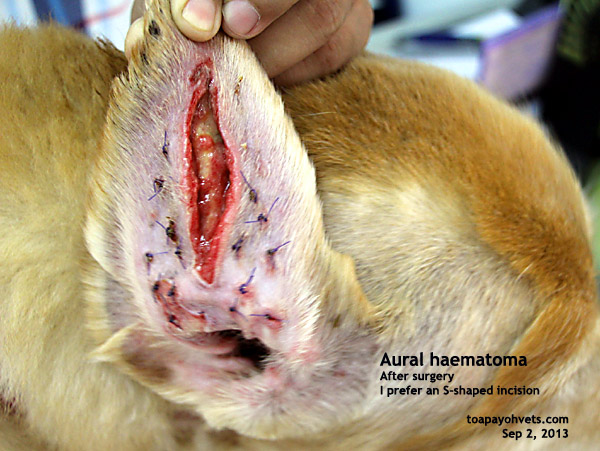
Veterinary and Travel Stories 1116. Aural haematoma surgery
HOPE Dog Rescue News From The Vacated Factory (2)

Ear new affection of ear and its treatment
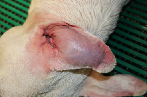
We kept putting cool compresses on it to sooth and applied moisturizing ointment to the flap as we found the skin looked a bit dried out due to being stretched out.
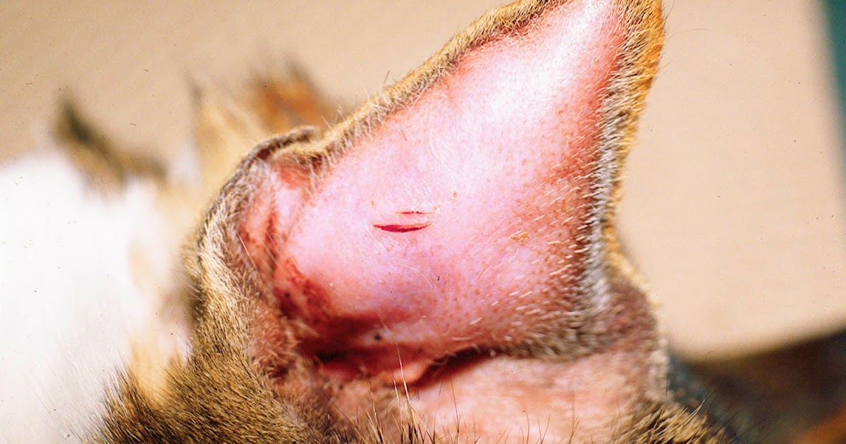
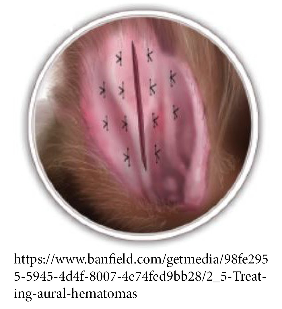
Aural hematoma cat after surgery. If the lesion is confined to just one part of the pinna, the swelling may be small. Ear hematomas are extremely painful & require prompt veterinary attention. The ear may or may not be bandaged after surgery.
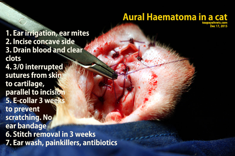
While under anesthesia, an incision is made along the length of the hematoma on the inner surface of the ear. When a blood vessel in the pinna breaks, it oozes fluid. The ear flap may partially or completely swell with blood.
An ear hematoma, or aural hematoma, is a collection of blood within the ear flap (pina). Subdermal hematomas/seromas form under the skin and are probably the most commonly type of hematoma or seroma. Ear hematoma, also called aural hematoma or auricular hematoma, is a common ear problem in cats.
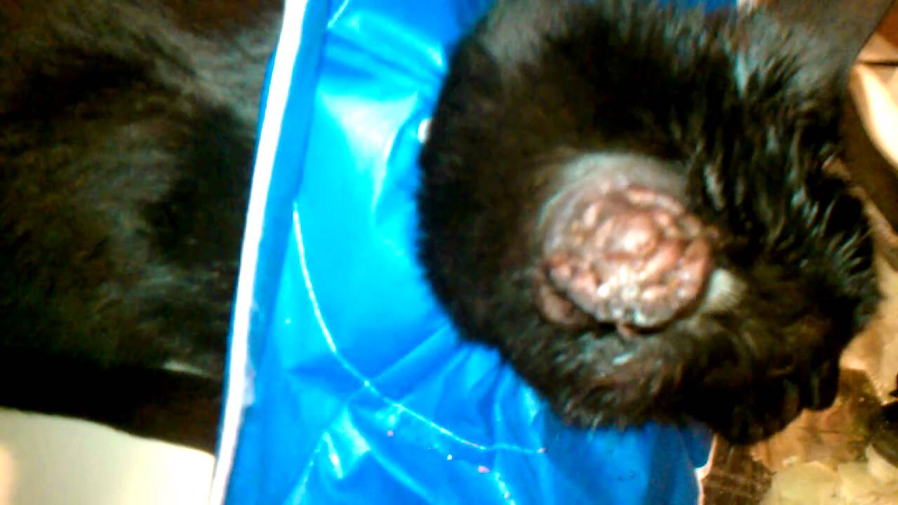
A 3 × 4 cm mass, macroscopically similar to an auricular hematoma, was visible on the convex surface and a smaller vascular lesion was present on the ear margin. The underlying causes include all conditions that result in otitis externa (infection of the external ear canal). Hematomas and seromas can occur anywhere in the body.
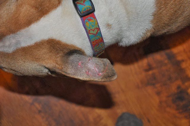
An aural (ear) hematoma is a collection of blood, serum, or a blood clot within the pinna (ear flap). This could be caused by ear mites, ticks, fleas, allergies, otitis externa (infection of the external ear canal). An ear hemotoma in dogs is the collection of blood between the dog's skin and the ear cartilage.
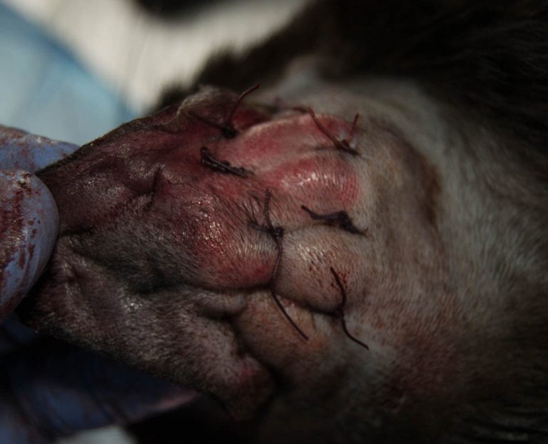
His was much bigger as well, taking up almost the entire ear flap. Eventually, the hematoma may become firm and thickened, resulting in a deformed “cauliflower” appearance. A hematoma is described as a mass of blood, liquid or coagulated, within the walls of tissues or an organ.
47 HQ Images Aural Hematoma Cat Cost Feline Patients House of Chaos

Surgical Repair of an Aural Hematoma in a Cat — Tails of a Shelter Vet
Aural Haematoma Cat Surgery maternity photos

Aural haematoma in Guinea pig (Cavia porcellus) Di Giuseppe 2018
The Cat Hemi after Ear Surgery YouTube
Aural Hematoma Veterinarian in MONTGOMERY, AL Animal Hospital of

Haematoma Surgery maternity photos

Animal Clinic at Thorndale, p.c.Aural or Ear Hematoma
Ear Hematoma What Are They and How Do Pets Get Them? Pawsitively Pets

Aural hematoma surgery YouTube

Can i pop my cats hematoma. Can i pop my cats hematoma.

Ear Hematoma YouTube
Cat Ear Hematoma Natural Treatment 37 Unconventional But Totally
